Research
Most animals in this world have an external bilaterally symmetric body plan. In simple terms, this translates to having a body plan with similar shapes, sizes and positions of tissues (e.g. eyes, ears, entire muscle and skeletal system etc.) on the left and right sides of the body. How do tissues form with such exquisite symmetry bilaterally? Our lab is interested in understanding the mechanism by which similar shapes and sizes of tissues emerge on the two sides of the body despite the presence of unavoidable biological noise during embryo development. We use zebrafish embryos as a model system and undertake an interdisciplinary approach involving high resolution imaging, advanced image analysis and collaboration with theoretical physicists to unravel this long-standing problem in developmental biology. We investigate how symmetry emerges in three different stages of embryonic development - during gastrulation, somite formation and craniofacial development.
Emergence of symmetry in convergence-extension flows during gastrulation
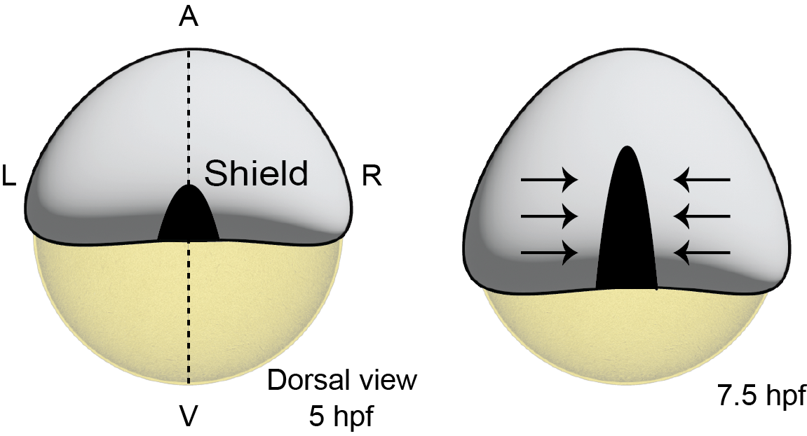
Early zebrafish embryos have a single body axis called the animal-vegetal (AV) axis. A few hours later (around 5 hours post fertilization (hpf)), a dorsal organizer (shield, in image below) forms establishing the dorsal-ventral (DV) axis of the embryo. Given two body axes (AV and DV), the third body axis (left-right) can be automatically defined as the three axes are perpendicular to each other. A couple of hours after establishment of such a 3D coordinate system, cells from the left and right sides of the embryo begin flowing towards the dorsal midline (see right image and video below). As the cells flow they intercalate with each other, thus shrinking the body axis in the left-right direction, while simultaneously elongating the body axis in the AV direction. These flows called convergence-extension flows are prevalent throughout development and has been extensively studied across systems. Here, we are trying to investigate whether these bilateral flows are symmetric and how such large-scale symmetry emerges in a dynamic process.
Emergence of symmetry in tissue shape and size during somite formation
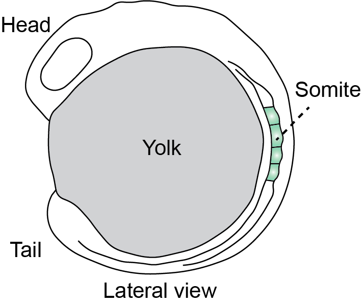
Somites, the precursors of the muscle and skeletal system, emerge with similar shapes and sizes on either side of the notochord during development. The movie below (taken with a multiview light-sheet microscope) with anterior of the embryo to the right, shows bilateral somite formation on either side of a tissue called the notochord. It was recently discovered that many somites form with imprecise shapes and sizes and the embryo performs error correction to ensure that symmetry emerges over time (Naganathan et al., 2022). The error correction process was investigated only from a tissue mechanics perspective and here we are interested in understanding the underlying cellular dynamics that drive tissue-scale shape changes in somites that ultimately ensure precise and bilaterally symmetric morphologies.
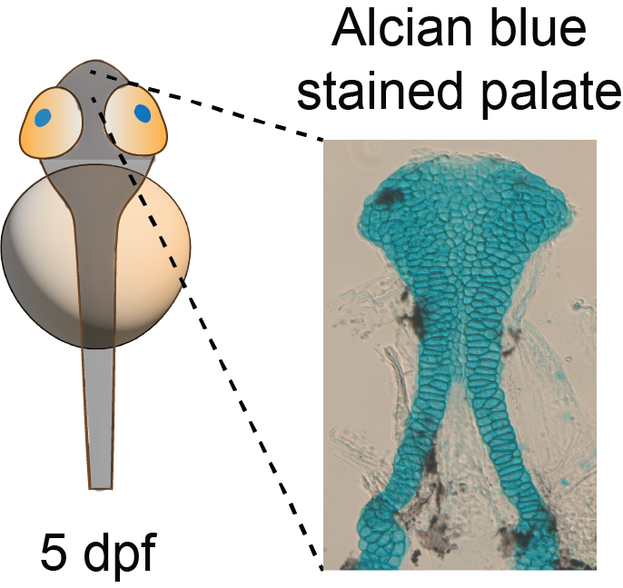
Emergence of symmetry in craniofacial features
Symmetry is highly evident in craniofacial features such as eyes, ears, palate and jaw. Most of the craniofacial cartilage and skeleton emerge from what are known as neural crest cells. These cells are first specified in the interface between the dorsal neural tube and the ectoderm, after which they undergo epithelial-mesenchymal transition and migrate large distances across the left and right sides to eventually reach the presumptive facial region. These bilateral migratory events are critical in craniofacial features emerging with precise shapes and sizes. Here, using the palate (roof of mouth) as a model system, we are investigating how bilateral flows drive formation of a symmetric palate along the facial midline.